Neurofluid imaging


Neurofluids is a novel umbrella term referring to all fluid-filled spaces in the brain. This includes the vasculature with arteries, arterioles, capillaries, venules, and veins as well as perivascular spaces (PVS), interstitial fluid (ISF), and the cerebrospinal fluid (CSF). Neurofluids have a profound effect on the brain’s function and physiology as well as being the origin of reserve mechanisms and the driver of waste clearance in the brain. With MRI we have the unique potential to image non-invasively the structure and function of neurofluids as well as associated downstream pathologies.
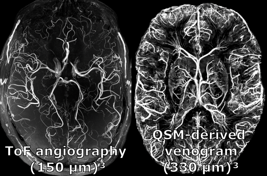
The quest to obtain ultra-high resolution vessel images resulted in some of the highest resolution MR images published to date with up to 140 µm isotropic resolution. The result show that with MRI it is feasible to image the pial arterial vasculature. Imaging pial arteries is crucial to understand the signal origin of fMRI as they show some of the largest responses to neuronal activity across all vessel types.
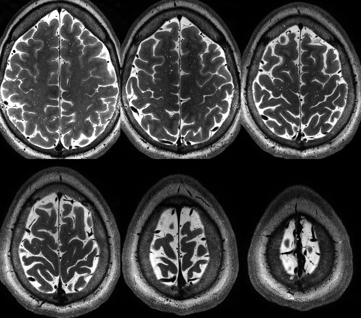
Similarly, multi-modal, ultra-high resolution MRI has been used to show that MR-visible PVS are associated with arteries and not veins in the basal ganglia.
Further, the integrity of (small) vessels and PVS is intertwined with diseases such as CSVD, ALS, and Alzheimer’s. Hence, imaging and assessing, e.g. with vessel distance mapping, them will improve our understanding of the diseases and, potentially, lead to new approaches for diagnostics and therapy.
Scientific contribution & selected publications
- Multi-modal study to show PVS are associated with arteries and not veins in the basal ganglia
Oltmer J, Mattern H, Beck J, Yakupov R, Greenberg S, Zwanenburg J, Arts T, Duzel E, van Veluw S, Schreiber S, Perosa V. Enlarged Perivascular Spaces in the Basal Ganglia are Associated with Arteries not Veins. Journal of Cerebral Blood Flow and Metabolism 2024 DOI:10.1177/0271678X241260629;
- Review on how the (micro)vasculature is impacting ALS disease progression. We discuss markers of how to assess the microvascular integrity with MRI (state of the art and promising new techniques) as well as how vascular supply patterns could play a role as a resistance or resilience mechanism in ALS.
Schreiber S, Bernal J, Arndt P, Schreiber F, Müller P, Morton L, Braun-Dullaeus RC, Valdés-Hernández MC, Duarte R, Wardlaw JM, Meuth SG, Mietzner G, Vielhaber S, Dunay IR, Dityatev A, Jandke S, Mattern H. *Brain Vascular Health in ALS Is Mediated through Motor Cortex Microvascular Integrity. Cells 2023 DOI:10.3390/cells12060957; *equal contribution
- Showing the advantage 7T and QSM over to detect microbleeds in CSVD patients.
Perosa V, Rotta J, Yakupov R, Kuijf HJ, Schreiber F, Oltmer JT, Mattern H, Heinze HJ, Düzel E, Schreiber S. Implications of quantitative susceptibility mapping at 7 Tesla MRI for microbleeds detection in cerebral small vessel disease. Frontiers in Neurology 2023 DOI: 10.3389/fneur.2023.1112312
- Case report: Leveraging high resolution MRI to study inflammation and counteractions in a CAA patient.
Schreiber S, John A-C, Werner C, Vielhaber S, Heinze H-J, Speck O, Würfel J, Behme D, Mattern H. Counteraction of inflammatory activity in CAA-related subarachnoid hemorrhage. Journal of Neurology 2022 DOI:10.1007/s00415-022-11437-9; *equal contribution
- Leveraging PMC to image pial arteries at 140 µm isotropic resolution.
Bollmann S, Mattern H, Bernier M, Robinson SR, Park D, Speck O, Polimeni JR. Imaging of the pial arterial vasculature of the human brain in vivo using high-resolution 7T time-of-flight angiography. eLife, 2022 DOI: 10.7554/eLife.71186
- Comparing pulsatility index in small arteries between CSVD patients and healthy controls leveraging the high resolution capabilities of 7T MRI.
Perosa V, Arts T, Assmann A, Mattern H, Speck O, Oltmer J, Heinze H-J, Düzel E, Schreiber S, Zwanenburg JJM. Pulsatility index in the basal ganglia arteries increases with age in elderly with and without cerebral small vessel disease. American Journal of Neuroradiology, 2022 DOI: 10.3174/ajnr.A7450
